Wenzhou virus 8 (WZV8) diagnosis by unique inclusions in shrimp hepatopancreatic E-cells and a molecular detection method
6 June 2022 | Jiraporn Srisala, Piyachat Saguanrut, Suparat Taengchaiyaphum, Rapeepun Vanichviriyakit, Kallaya Sritunyalucksana and Timothy F. Flegel | 2545 Downloads | .pdf | 894.71 KB | Health and Biosecurity, Shrimp
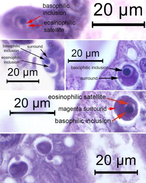
To assist shrimp pathologists worldwide we are providing here photomicrographs of unique basophilic inclusions that are produced by Wenzhou shrimp virus 8 (WZV8) (Li et al. 2015) that was discovered in 2015 by wide screening of marine animals for RNA viruses using high throughput sequencing (GenBank record KX883984.1). A more recent publication from China (Liu et al. 2021) also gives the full sequence under GenBank record OK662577 that is highly similar to WZV8 (97% coverage and 95.4% sequence identity) but under the newly proposed name Penaeus vannamei picornavirus (PvPV). Although that paper contained no histological analysis, it did include an electron micrograph of a cytoplasmic viral inclusion within a vacuole of an unspecified hepatopancreatic epithelial cell type (Liu et al., 2021).
Using the sequence of KX883984.1, we designed PCR primers and in situ hybridization probes for detection of WZV8. Subsequent ISH assays with shrimp RT-PCR positive for WZV8 samples allowed us to identify unique inclusions described herein as linked to WZV8 in hematoxylin and eosin (H&E) stained tissues. In some of the specimens positive for WZV8 with ISH assays, positive ISH reactions were also seen in normal nuclei in the central region of the HP and in the subcuticular epithelium and underlying connective tissue (especially in the stomach) indicating that these tissues are of no use for histological diagnosis of WZV8 infection because of their normal appearance with H&E staining.
Going back over our previous histological reports and archived slides, we have found the unique WZV8 inclusions in E-cells of normal shrimp samples from several shrimp farming countries in Austral-Asia since at least 2008. More recently we have obtained samples of P. vannamei from the Americas that also show these inclusions. We have noticed these inclusions for many years as unique basophilic, cytoplasmic inclusions of unknown origin that occur mostly in E-cells of the tubule epithelia of the hepatopancreas (HP) of both diseased and normal, cultivated P. monodon and P. vannamei. In diseased samples, mortality was ascribed to bacteria or known lethal viruses. As a result, the additional presence of these inclusions of unknown origin and their relatively common presence also in shrimp with no signs of disease resulted in their relative neglect while efforts were focused on more urgent problems.
We urge shrimp pathologists to review their records and archived and current specimens for the presence of the unique WZV8 E-cell inclusions described herein. Hopefully, this will result in data that will provide a global view of the current prevalence and impact of WZV8-like infections.
Copyright, all rights reserved.

