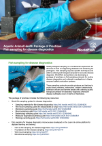
Quick fish sampling for disease diagnostics: Histology sampling guide
30 January 2024 | Delamare-Deboutteville, J., Ali S.E.S.M., and Chadag V.M. | 213 Downloads | .pdf | 4.3 MB | Health and Biosecurity
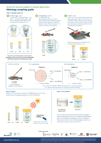
During investigation of an abnormal mortality event, fish tissues (or biopsies) for histopathology analysis are collected for disease diagnosis. Histology consists in the preparation of thin, stained tissue sections for microscopic examination to study their structure and function.
Histopathology is the study of disease and disease processes by looking at the change in the anatomy or anomalies from cells, tissues and organs as seen through a microscope.
WorldFish and partners developed this quick fish-sampling guide for histology. Standard biopsy specimens for histological examination consists of fixed sections of brain, gill, heart, intestine, kidney, liver and spleen. Other tissues may be collected in the presence of external lesions/ulcers (e.g., eye, skin, muscle).
A free online training course on histology sampling is also available via Learn.ink.
Publisher: WorldFish
Rights: Creative Commons Attribution-NonCommercial.